
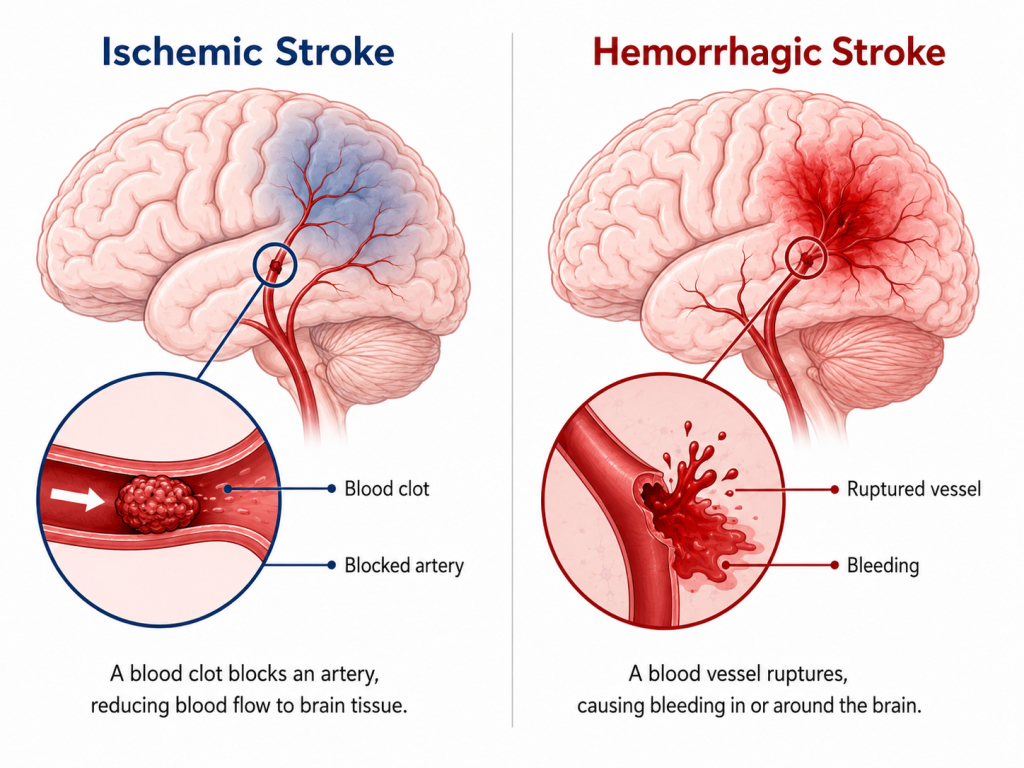
A stroke occurs when blood flow to part of the brain is interrupted. The two main types of stroke are ischemic stroke, caused by a blocked blood vessel, and hemorrhagic stroke, caused by bleeding from a ruptured blood vessel.
Ischemic stroke is the more common type. It occurs when an artery supplying the brain becomes blocked, reducing or stopping blood flow to brain tissue. The term acute ischemic stroke refers to the early phase of the event, particularly during emergency assessment and treatment.
Without adequate blood flow, affected brain tissue does not receive the oxygen and nutrients it needs, and ischemic injury can progress over time. Rapid assessment is therefore important because some treatments for acute ischemic stroke are time-dependent.
Treatment is not the same for every patient. Clinical assessment and imaging are used to determine the type of stroke, identify the location and extent of an arterial blockage, and assess which treatments may be appropriate.
What Is Acute Ischemic Stroke?
An acute ischemic stroke occurs when blood flow through an artery supplying the brain is interrupted by a blockage.
The effects depend on factors such as the part of the brain affected, the artery involved and the extent of reduced blood flow. Symptoms and the severity of neurological impairment can therefore vary considerably between patients.
Within the affected area, some brain tissue may sustain irreversible injury while surrounding tissue with reduced blood flow may remain viable for a period of time. The rate at which ischemic injury progresses varies between individuals and can be influenced by factors such as the location of the blockage and collateral blood flow.
This is why suspected acute ischemic stroke requires urgent medical assessment.
What Causes Ischemic Stroke?
Ischemic stroke occurs when an artery supplying the brain becomes blocked. The blockage can develop in different ways.
In some cases, a blood clot forms within an artery that has become narrowed or affected by atherosclerosis.
In other cases, a clot forms elsewhere in the circulation and travels until it blocks an artery supplying the brain. This is known as an embolic stroke.
One form of embolic ischemic stroke is cardioembolic stroke, in which the clot originates in the heart. Atrial fibrillation is an important cause because this abnormal heart rhythm can allow blood clots to form in the heart and subsequently travel through the circulation to the brain.
Ischemic stroke can also result from disease affecting smaller cerebral blood vessels, arterial dissection and other less common causes. In some patients, a definite cause cannot be identified even after investigation.
Understanding the likely cause is important because it can influence how clinicians approach the prevention of another stroke.
Risk Factors for Ischemic Stroke
A range of cardiovascular, metabolic and lifestyle factors are associated with an increased risk of ischemic stroke.
Important risk factors include:
- High blood pressure
- Atrial fibrillation
- Diabetes
- High cholesterol and other lipid disorders
- Smoking
- Physical inactivity
- Overweight and obesity
- Cardiovascular disease
- Increasing age
Some risk factors, such as age, cannot be changed. Others may be modified through lifestyle measures, medical treatment or both.
The contribution of individual risk factors varies between people, so approaches to stroke prevention should be based on individual clinical circumstances.
What Are the Symptoms of Acute Ischemic Stroke?
Acute ischemic stroke can cause sudden neurological symptoms. However, many of the same symptoms can also occur with hemorrhagic stroke. Symptoms alone cannot reliably determine which type of stroke has occurred, which is why urgent clinical assessment and imaging are important.
Possible stroke symptoms include:
- Sudden weakness or numbness of the face, arm or leg, particularly on one side of the body
- Sudden difficulty speaking or understanding speech
- Sudden confusion
- Sudden loss of balance or coordination
- Sudden difficulty walking
- Sudden changes in vision in one or both eyes
- Sudden dizziness
The American Stroke Association uses the B.E. F.A.S.T. mnemonic to help people recognise several common stroke warning signs:
- B – Balance: Sudden loss of balance or coordination
- E – Eyes: Sudden vision changes
- F – Face: Facial weakness or drooping
- A – Arm: Arm weakness or numbness
- S – Speech: Difficulty speaking or understanding speech
- T – Time: Seek emergency medical help
These warning signs apply to suspected stroke generally rather than only to ischemic stroke because the type of stroke cannot reliably be determined from symptoms alone.
What If Stroke-Like Symptoms Disappear?
Stroke-like symptoms may sometimes resolve before a person receives medical assessment. This can occur with a transient ischemic attack (TIA).
A TIA involves temporary neurological dysfunction caused by reduced blood flow to the brain without acute infarction. It is therefore not the same as an ischemic stroke, although the symptoms can be similar.
The distinction cannot necessarily be made when symptoms first occur. A TIA also requires urgent medical assessment because it is associated with an increased risk of subsequent stroke, particularly during the first few days.
How Acute Ischemic Stroke Is Diagnosed
When a patient presents with suspected stroke, clinicians first need to determine whether a stroke has occurred, assess the neurological deficit and distinguish an ischemic stroke from intracranial bleeding or another condition that can produce similar symptoms.
Initial assessment may include a neurological examination, relevant medical history, measurement of vital signs such as blood pressure, and point-of-care blood glucose testing.
A structured neurological assessment such as the National Institutes of Health Stroke Scale (NIHSS) may be used to assess the severity of neurological deficits. The NIHSS contributes to the overall clinical assessment but does not by itself determine which treatment a patient should receive.
Imaging Tests Used in Acute Stroke Assessment
Imaging is an essential part of acute stroke assessment. Different imaging techniques provide different information about the brain and its blood vessels.
A noncontrast CT scan is commonly used as an initial imaging examination because it can rapidly identify intracranial hemorrhage. Early ischemic changes may also be visible, although an acute ischemic stroke may not always be apparent on the initial CT scan.
CT angiography or MR angiography can be used to examine arteries supplying the brain and identify an arterial occlusion. This information is particularly relevant when mechanical thrombectomy is being considered.
MRI can provide additional information about brain tissue and ischemic injury and may be used in selected patients.
Perfusion imaging assesses patterns of blood flow within the brain. It may be used to support treatment selection in certain situations, including some patients who present later or whose time of stroke onset is uncertain.
Not every patient requires every type of imaging. The imaging approach depends on the clinical circumstances, and additional imaging should not unnecessarily delay time-sensitive treatment in otherwise eligible patients.
How Acute Ischemic Stroke Is Treated
Treatment depends on factors including the patient’s neurological symptoms, time of symptom onset or last known well, imaging findings, location of the arterial blockage, medical history and other clinical considerations.
Two important treatments aimed at restoring blood flow are intravenous thrombolysis and mechanical thrombectomy.
They are used in different clinical circumstances and are not necessarily alternatives. Some eligible patients may receive intravenous thrombolysis followed by mechanical thrombectomy.
Intravenous Thrombolysis for Acute Ischemic Stroke
Intravenous thrombolysis is a treatment in which a clot-dissolving medication is given through a vein to eligible patients with acute ischemic stroke.
Current guidelines recommend alteplase or tenecteplase for eligible adults presenting within 4.5 hours of symptom onset or the time they were last known to be well.
Eligibility depends on more than time alone. Clinicians consider factors including the neurological deficit, imaging findings, medical history and contraindications to thrombolysis.
Selected patients who present beyond the conventional treatment window, or whose exact time of stroke onset is unknown, may also be considered for intravenous thrombolysis when specific clinical and imaging criteria are met.
These extended treatment approaches do not mean that time is unimportant. Eligible patients should still be assessed and treated as rapidly as possible.
Mechanical Thrombectomy
Mechanical thrombectomy is a catheter-based procedure used to remove an obstructing clot from an artery supplying the brain.
It is an established treatment for selected patients with acute ischemic stroke caused by an eligible large-vessel occlusion, in which one of the larger arteries supplying the brain is blocked.
During the procedure, a catheter is introduced into the arterial system and guided to the site of the blockage using imaging. The clot may then be removed using a stent retriever, an aspiration system or a combination of techniques.
Randomised clinical trials have shown improved functional outcomes with mechanical thrombectomy compared with medical management alone in appropriately selected patients with large-vessel occlusion.
Eligibility depends on factors including the artery involved, neurological findings, imaging findings, time since symptom onset or last known well, baseline function and other clinical considerations.
Why Treatment Timing Matters
When an artery supplying the brain becomes blocked, ischemic injury can progress over time.
Within the affected region, some brain tissue may already have sustained irreversible injury while other tissue with reduced blood flow may remain potentially viable. One aim of reperfusion treatment is to restore blood flow before potentially salvageable tissue becomes irreversibly damaged.
The rate at which ischemic injury progresses differs between patients. Treatment decisions therefore consider time together with clinical and imaging findings, rather than relying on time alone in every situation.
Nevertheless, intravenous thrombolysis and mechanical thrombectomy remain time-sensitive treatments. Rapid recognition of stroke symptoms, activation of emergency medical services and efficient hospital assessment can reduce delays in determining the appropriate treatment pathway.
Reducing the Risk of Ischemic Stroke
Reducing the risk of ischemic stroke involves identifying and managing modifiable risk factors.
Depending on the individual, this may include:
- Managing high blood pressure
- Managing diabetes and lipid disorders
- Stopping smoking
- Maintaining regular physical activity
- Following an appropriate healthy dietary pattern
- Managing atrial fibrillation and other relevant cardiovascular conditions
- Taking preventive medications when clinically indicated
The approach differs between people who have never experienced a stroke and those who have previously had an ischemic stroke or TIA.
After an ischemic stroke, further investigation may help identify the likely cause. Secondary prevention can then be tailored to the underlying stroke mechanism and the individual’s other cardiovascular risk factors.
Acute Ischemic Stroke: Why Rapid Assessment Matters
Acute ischemic stroke is a medical emergency, but diagnosis and treatment are not the same for every patient.
Because ischemic and hemorrhagic strokes can produce similar symptoms, clinical assessment and imaging are needed to determine the type of stroke and guide treatment decisions.
For eligible patients with acute ischemic stroke, intravenous thrombolysis and mechanical thrombectomy are established reperfusion treatments used in different clinical circumstances. Treatment decisions depend on timing together with clinical and imaging findings.
Recognising possible stroke symptoms and seeking emergency medical care promptly allows assessment and, where appropriate, treatment to begin as early as possible.
Clinical References
- Prabhakaran S, Gonzalez NR, Zachrison KS, et al. 2026 Guideline for the Early Management of Patients With Acute Ischemic Stroke: A Guideline From the American Heart Association/American Stroke Association. Stroke. 2026;57(8):e316–e436. doi:10.1161/STR.0000000000000513. Corrected July 2026.
- American Stroke Association. Types of Stroke and Treatment.
- American Stroke Association. Ischemic Stroke (Clots).
- American Stroke Association. Stroke Symptoms and Warning Signs.
- Amin HP, Madsen TE, Bravata DM, et al. Diagnosis, Workup, Risk Reduction of Transient Ischemic Attack in the Emergency Department Setting: A Scientific Statement From the American Heart Association. Stroke. 2023;54:e109–e121. doi:10.1161/STR.0000000000000418.
- Bushnell C, Kernan WN, Sharrief AZ, et al. 2024 Guideline for the Primary Prevention of Stroke: A Guideline From the American Heart Association/American Stroke Association. Stroke. 2024;55(12):e344–e424. doi:10.1161/STR.0000000000000475.
- Kleindorfer DO, Towfighi A, Chaturvedi S, et al. 2021 Guideline for the Prevention of Stroke in Patients With Stroke and Transient Ischemic Attack: A Guideline From the American Heart Association/American Stroke Association. Stroke. 2021;52:e364–e467. doi:10.1161/STR.0000000000000375.
This article is intended for general educational purposes and does not replace individual medical advice, diagnosis or treatment from a qualified healthcare professional.