
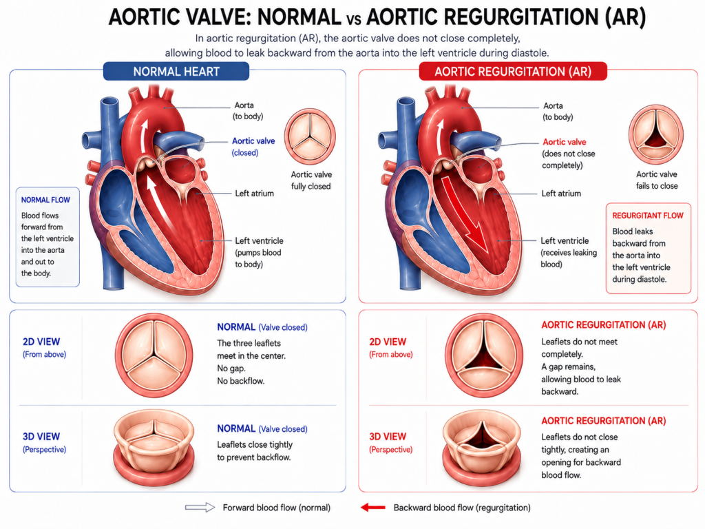
The heart has four valves that help keep blood moving in the right direction. One of these is the aortic valve, which sits between the left ventricle, the heart’s main pumping chamber, and the aorta, the body’s main artery.
Aortic regurgitation, also called aortic valve regurgitation, occurs when the aortic valve does not close completely. This allows some blood to flow backwards from the aorta into the left ventricle instead of continuing forward to the rest of the body.
Over time, this backward flow increases the volume of blood the left ventricle must handle. In chronic aortic regurgitation, the heart can gradually adapt, and some people may have no symptoms for many years. As the condition progresses, however, the left ventricle may enlarge and its pumping function may eventually decline.
Aortic regurgitation can also develop suddenly. Acute severe aortic regurgitation is a medical emergency because the heart has little time to adapt to the sudden increase in blood volume.
Understanding the causes, symptoms, diagnosis and treatment of aortic regurgitation can help patients better understand the condition and why ongoing assessment may be important.
What Is Aortic Regurgitation?
Aortic regurgitation is a type of heart valve disease in which the aortic valve does not close properly after blood has been pumped from the left ventricle into the aorta.
The resulting backward flow of blood places an additional volume load on the left ventricle.
Aortic regurgitation may be:
- Chronic, developing gradually over time
- Acute, developing suddenly as a result of damage to the valve or aorta
In chronic aortic regurgitation, the left ventricle can gradually enlarge as it adapts to the increased blood volume. This helps explain why some people may remain symptom-free even when the regurgitation is significant.
In acute aortic regurgitation, there is little or no time for this adaptation to occur. Severe acute regurgitation can lead to rapid deterioration, including pulmonary oedema, low blood pressure and heart failure. Urgent specialist assessment is required, and emergency surgery may be necessary depending on the underlying cause.
What Causes Aortic Valve Regurgitation?
Aortic regurgitation can result from abnormalities of the valve leaflets themselves or from changes in the aortic root or ascending aorta that prevent the valve from closing normally.
Causes may include:
- Bicuspid aortic valve
- Degenerative changes affecting the aortic valve
- Enlargement of the aortic root or ascending aorta
- Connective tissue disorders
- Infective endocarditis
- Rheumatic heart disease
- Aortic dissection
- Trauma affecting the valve or aorta
A bicuspid aortic valve is a congenital condition in which the valve has two leaflets instead of the usual three. This difference in valve structure can affect how the valve functions and may contribute to aortic regurgitation or other aortic valve disease over time.
Aortic regurgitation may also develop when the aortic root or ascending aorta becomes enlarged. As the surrounding anatomy changes, the valve leaflets may no longer meet properly when the valve closes, allowing blood to leak backwards.
Acute aortic regurgitation is more commonly associated with conditions that suddenly damage the valve or aorta, such as infective endocarditis or aortic dissection.
Is Aortic Regurgitation Serious?
The significance of aortic regurgitation depends on factors such as its severity, how quickly it develops and how it affects the left ventricle.
Mild aortic regurgitation may require monitoring without immediate treatment. More severe chronic aortic regurgitation can gradually increase the workload on the heart, causing the left ventricle to enlarge and, over time, potentially affecting its ability to pump effectively.
Importantly, these changes can begin before symptoms become noticeable. This is why regular assessment may be recommended even when a person feels well.
Acute severe aortic regurgitation is different. Because the heart has no time to adapt to the sudden increase in blood volume, the condition can become life-threatening and requires urgent medical assessment.
What Are the Symptoms of Aortic Regurgitation?
Many people with chronic aortic regurgitation have few or no symptoms during the early stages.
Because the left ventricle can adapt gradually to the additional blood volume, significant regurgitation may be present before symptoms become noticeable.
As the condition progresses, symptoms may include:
- Shortness of breath, particularly during physical activity
- Fatigue
- Reduced exercise tolerance
- Awareness of a forceful or irregular heartbeat
- Chest pain or discomfort in some patients
- Shortness of breath when lying flat or during the night
- Swelling of the ankles or legs in more advanced heart failure
Symptoms of chronic aortic regurgitation often develop gradually.
In acute severe aortic regurgitation, patients may develop sudden severe shortness of breath, pulmonary oedema, low blood pressure or other signs of rapidly worsening heart function. This requires urgent medical assessment.
Aortic regurgitation may also be identified before symptoms develop, for example when a healthcare professional detects a characteristic heart murmur during a physical examination.
How Is Aortic Regurgitation Diagnosed?
Aortic regurgitation may first be suspected based on symptoms, medical history or findings during a physical examination.
The main imaging test used to evaluate suspected aortic regurgitation is transthoracic echocardiography (TTE).
Echocardiography
An echocardiogram uses ultrasound to create moving images of the heart and its valves.
In aortic regurgitation, echocardiography can help clinicians assess:
- The structure and movement of the aortic valve
- The presence and severity of regurgitation
- The possible mechanism causing the regurgitation
- The size of the left ventricle
- How well the left ventricle is pumping
- The dimensions of the aortic root and ascending aorta
The severity of aortic regurgitation is assessed by considering multiple echocardiographic findings together rather than relying on a single measurement.
Other Imaging Tests
Additional imaging may be recommended when more information is needed.
Transoesophageal echocardiography (TEE)
TEE can provide more detailed images of the heart and may be used when transthoracic imaging does not provide sufficient information or when more detailed assessment of valve anatomy is required.
Cardiac magnetic resonance imaging (CMR)
CMR can provide detailed measurements of heart chamber size and function and can help quantify regurgitant blood flow. It may be useful when echocardiographic findings are inconclusive or when additional assessment is needed.
Cardiac computed tomography (CT)
Cardiac CT can provide detailed information about the aortic root, ascending aorta and surrounding anatomy. It may also form part of anatomical assessment and planning before certain procedures.
Why Is Regular Follow-Up Important?
People with chronic aortic regurgitation may feel well even while changes are occurring in the heart.
For this reason, patients with known aortic regurgitation may require ongoing clinical review and repeat echocardiography. The frequency of follow-up depends on factors such as the severity of the regurgitation, symptoms and changes in heart size or function.
Follow-up allows healthcare professionals to monitor:
- Changes in symptoms
- Progression of the regurgitation
- Changes in left ventricular size
- Changes in left ventricular pumping function
- Changes in the aortic root or ascending aorta
This is particularly important in severe chronic aortic regurgitation because valve intervention may sometimes be considered based on changes seen on imaging before significant symptoms develop.
How Is Aortic Regurgitation Treated?
Treatment depends on the severity and cause of the regurgitation, its effect on the heart and the individual patient’s clinical circumstances.
Not everyone with aortic regurgitation requires immediate valve intervention. Treatment may involve regular monitoring, management of associated conditions or intervention on the aortic valve or aorta.
Monitoring and Medical Treatment
People with mild or moderate aortic regurgitation who have no symptoms may be managed with regular follow-up rather than immediate intervention.
Medication may be used to manage associated conditions such as high blood pressure or, when appropriate, symptoms of heart failure.
However, medication does not correct the underlying structural problem causing aortic regurgitation. In severe disease, valve intervention may be required when established clinical and imaging criteria are met.
When Is Valve Intervention Considered?
For patients with severe aortic regurgitation, the decision to intervene is based on more than symptoms alone.
Healthcare professionals consider factors including:
- Whether symptoms are present
- Left ventricular size
- Left ventricular pumping function
- Changes seen on follow-up imaging
- Enlargement of the aortic root or ascending aorta
- The underlying cause of the regurgitation
- Other cardiac conditions
- The patient’s overall health and procedural risk
Clinical guidelines use established measurements of left ventricular size and function to help determine when intervention should be considered.
This is important because chronic aortic regurgitation can cause progressive changes in the left ventricle before a patient feels unwell. In some patients with severe aortic regurgitation, intervention may therefore be recommended before symptoms develop if imaging shows significant enlargement of the left ventricle or a decline in its pumping function.
For patients being considered for valve intervention, assessment may involve a multidisciplinary Heart Team, bringing together specialists such as cardiologists, cardiac surgeons, imaging specialists and interventional cardiologists.
The aim is to determine whether intervention is needed and, if so, which treatment approach is most appropriate for the individual patient.
Treatment Options for Severe Aortic Regurgitation
When valve intervention is required, the appropriate approach depends on the cause of the aortic regurgitation, the anatomy of the valve and aorta, the patient’s overall health and other clinical factors.
Aortic Valve Repair
In selected patients, the patient’s own aortic valve may be suitable for repair rather than replacement.
Whether repair is possible depends on factors such as the cause of the regurgitation, valve anatomy, tissue quality and the condition of the aortic root.
Valve repair or valve-sparing surgery may also be considered in selected patients who require surgery for enlargement of the aortic root or ascending aorta. These procedures require appropriate anatomy and relevant surgical expertise.
Surgical Aortic Valve Replacement
Surgical aortic valve replacement (SAVR) remains an established treatment for patients with severe aortic regurgitation who require valve replacement and are suitable for surgery.
During surgery, the diseased aortic valve is replaced with a prosthetic valve.
Treatment planning may take into account factors such as:
- Age
- Overall health
- Valve and aortic anatomy
- Other heart conditions
- Surgical risk
- The choice between mechanical and biological replacement valves
- Long-term treatment considerations
In some patients, surgery on the aortic root or ascending aorta may be required at the same time.
Transcatheter Aortic Valve Replacement
Transcatheter aortic valve replacement (TAVR or TAVI) allows a replacement valve to be implanted using a catheter-based approach.
Transcatheter treatment is well established for many patients with aortic stenosis, but its role in isolated aortic regurgitation is more limited.
Aortic regurgitation can present different anatomical challenges from aortic stenosis. For example, some patients have enlargement of the aortic root or valve opening and little or no calcification of the native valve. These characteristics can affect the positioning and stability of some transcatheter valves.
Transcatheter treatment may be considered for carefully selected patients with severe aortic regurgitation who are not suitable candidates for surgery, provided the anatomy is appropriate.
Suitability also depends on the individual patient’s clinical circumstances, the technology being used and applicable regulatory indications. As evidence and available technologies continue to develop, treatment decisions require individual assessment by an appropriately experienced clinical team.
Conclusion
Aortic regurgitation occurs when the aortic valve does not close completely, allowing blood to flow backwards from the aorta into the left ventricle.
Chronic aortic regurgitation may develop gradually and remain symptom-free for many years. Over time, however, the additional volume of blood can cause the left ventricle to enlarge and may eventually affect heart function. Acute severe aortic regurgitation develops suddenly and is a medical emergency.
Echocardiography is the main imaging test used to diagnose and assess aortic regurgitation, with CMR, CT or transoesophageal echocardiography providing additional information when needed.
Treatment depends on the severity and cause of the condition, symptoms, changes in the heart and the individual patient’s clinical circumstances. Some patients require regular monitoring, while others may require aortic valve repair or replacement. Surgical treatment remains central to the management of severe aortic regurgitation when intervention is required, while transcatheter treatment may be an option for carefully selected patients who are not suitable for surgery.
Regular clinical and imaging follow-up is important because changes in the heart can occur before symptoms become obvious.
Clinical References:
- 2025 ESC/EACTS Guidelines for the Management of Valvular Heart Disease
- 2020 ACC/AHA Guideline for the Management of Patients With Valvular Heart Disease
This article is intended for general educational purposes and does not replace individual medical advice, diagnosis or treatment from a qualified healthcare professional.